
Cone Beam
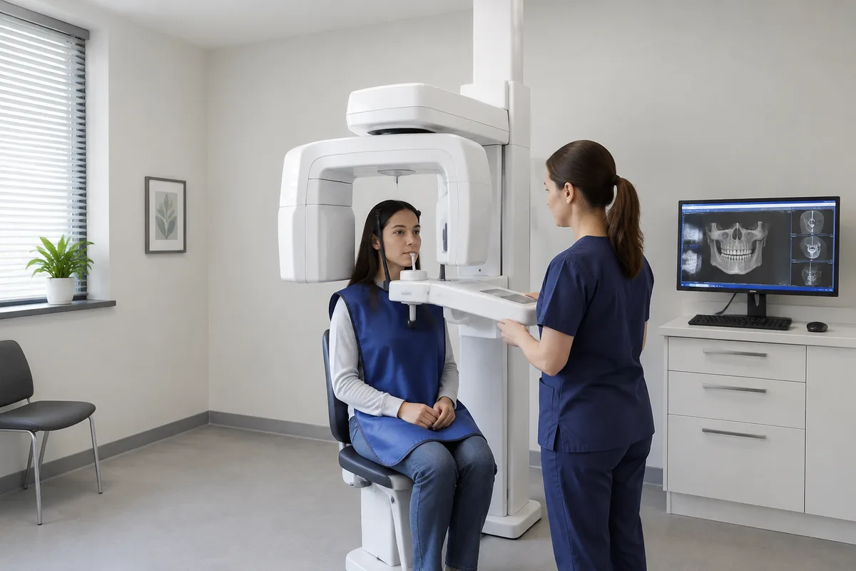
Cone Beam
Cone beam computed tomography, often shortened to CBCT, is an imaging technology that creates three-dimensional (3D) images of the structures inside your mouth and jaw. At Sam's Smile Dental, cone beam imaging is one of the on-site facilities that gives our team a fuller picture of what is happening beneath the surface.
What cone Beam is
A traditional dental X-ray is a flat, two-dimensional picture. It is useful for many things, but it compresses depth, so structures can overlap and hide one another. A cone beam scan works differently. The machine rotates around your head and captures many images from different angles using a cone-shaped X-ray beam. Software then combines those images into a single 3D model that can be viewed from any direction and in thin slices.
That 3D view lets a dentist look at teeth, roots, jawbone, nerve pathways, and the spaces around them without anything overlapping. Cone beam technology was developed as an improvement on two older approaches: flat X-rays, which lack depth, and conventional medical CT scans, which are larger machines built for the whole body.
Common things a cone beam image can help show include:
- Bone volume and shape — how much bone is present in the jaw and how it is contoured.
- Root anatomy — the number, length, and curvature of tooth roots.
- Nerve and sinus locations — where important structures sit relative to the teeth.
- Impacted teeth — the position of teeth that have not erupted normally.
- Airway space — the shape of the passages behind the mouth and nose.
How we use it at Sam’s Smile Dental
Our cone beam unit is listed among the practice's on-site facilities. It produces 3D images, which give our team more information than a flat image when a closer look is needed.
Cone beam imaging is generally used in areas such as dental implants, root canal therapy, tooth extraction and wisdom teeth removal, and sleep-related concerns. Sam's Smile Dental offers all of these services. Whether a scan is appropriate depends on your situation, and your dentist will decide that after an exam and a conversation about your needs.
The practice also has other diagnostic and treatment technology on site, including laser cavity detection, laser dentistry, and Isolite. Each has its own purpose. Cone beam is the one that shows structures in three dimensions, while the others address cavity detection, treatment, and comfort during procedures.
What it means for you
For patients, the main value of a 3D scan is clarity. When your dentist can see the whole picture, there is less guesswork and more to base a recommendation on. In general, cone beam imaging offers these benefits:
- Better planning — seeing bone, roots, and nerves in three dimensions helps when a procedure needs precision.
- Clearer explanations — a 3D image is easier to understand than a flat one, so you can see what your dentist is describing.
- Fewer surprises — hidden anatomy or unusual root shapes can be identified before treatment begins.
- Convenience — a scan can often be done in the dental setting rather than at a separate imaging center, which may save you a trip.
A scan will not replace a thorough exam. It is one tool that works together with your dentist's evaluation and your own goals.
What to expect during your visit
Before. Your team will explain why a scan is being recommended and answer your questions. Preparation is generally minimal. You may be asked to remove glasses, jewelry, hearing aids, or removable dental appliances, because metal can interfere with the image. Let the team know if you are or might be pregnant, so they can talk through your options.
During. Most cone beam units have you stand or sit while the scanner rotates around your head. You will be asked to stay still and may be given a bite guide or head support to steady you. The rotation itself generally lasts well under a minute, and the machine does not touch you. Most people feel nothing during the scan.
After. The images appear on a screen almost immediately. Your dentist can review them with you, point out relevant areas, and explain how they affect your care. You can usually go about your day with no recovery. If you want to know how long your particular visit will take, ask our team when you schedule.
Frequently asked questions
Is a cone beam scan safe?
Cone beam imaging uses X-rays, so it involves radiation exposure. Doses are generally lower than those of a conventional medical CT scan, and dental teams follow the principle of using imaging only when it will help answer a clinical question. If you have concerns, ask your dentist why a scan is recommended for you.
Does it hurt?
No. The scan is non-invasive, and nothing is placed inside your mouth beyond a bite guide in some cases. You simply hold still while the machine moves around you. People who feel uneasy in tight spaces can mention it beforehand, since the unit is open around the head rather than a closed tube.
How long does it take, and how often is it needed?
The scan itself generally takes less than a minute, though positioning and review add a little time. It is not a routine part of every dental visit. It is typically used when a flat X-ray cannot give enough detail, such as when planning certain procedures. Your dentist will advise whether it makes sense for you.
Who is cone beam imaging suitable for?
Many adults and some older children can have a scan when a dentist finds it useful. It may not be recommended during pregnancy unless clearly necessary, and your dentist will weigh the benefits against the alternatives. Talk with our team about your health history so they can recommend what fits you.
If you would like to learn more about cone beam imaging, or wonder whether it could help with your care, we invite you to book a visit or get in touch with Sam’s Smile Dental. We are happy to answer your questions.
This content is for general information only and isn't dental or medical advice. Talk with your dentist or orthodontist about your own situation. If you have a medical emergency, call 911.